Electrophysiology CINRE, hospital BORY
Atrial Fibrillation: Guidelines (2026) Compendium / 2.3 Trigger and Substrate of Atrial Fibrillation

Trigger and Substrate of Atrial Fibrillation


Atrial remodelling is a consequence of atrial cardiomyopathy,

  • which develops on the basis of risk factors, comorbidities, and genetics.

In remodelled atria, electrical changes develop that create the substrate for atrial fibrillation (AF).

Electrical changes arise mainly at sites where one anatomical structure transitions into another:

  • Most commonly at the ostia (antra) of the pulmonary veins, where the pulmonary veins connect to the left atrium.
  • Specifically, these are myocardial sleeves extending 1–4 cm into the pulmonary veins.
    • This represents atrial myocardium extending through the ostia into the interior of the pulmonary veins.

An episode of AF requires two components for initiation and maintenance:

  • Trigger – initiates the episode of AF.
  • Substrate – maintains the episode of AF.
Diagram of atrial fibrillation illustrating trigger sites and the atrial substrate, including pulmonary vein ostia, the posterior wall, and other anatomical regions.

Trigger

  • A focally electrically remodelled region in the atria, most commonly located at the pulmonary vein ostia.
  • Foci generate electrical impulses, resulting in atrial arrhythmias (premature atrial contractions, salvos, atrial tachycardia <30 s).
    • Premature atrial contractions are most common.
  • Premature atrial contractions arising from these foci may be isolated or occur in salvos (3–5 premature beats).
  • These premature atrial contractions may initiate AF if a suitable substrate is present.

Substrate

  • An electrically remodelled region in the atria, most commonly located at the pulmonary vein ostia.
  • The substrate ≠ the trigger, although they may be located in close proximity.
  • The substrate does not spontaneously generate premature atrial contractions.
  • The substrate is activated by electrical activity from the foci (premature atrial contractions).
  • After activation, the substrate begins to generate electrical impulses at a rate of 300–600/min, seen on the ECG as an episode of AF.

The initiation and duration of an AF episode depend on the complex interaction between trigger, substrate, and autonomic modulation.

  • The more advanced the substrate, the smaller the trigger required.

Which premature atrial contraction or salvo activates the substrate and how long the AF episode persists is extremely variable. It depends mainly on:

  • The extent and localization of the atrial substrate (fibrosis, left atrial dilatation).
  • Electrophysiological properties of the atrial myocardium (refractoriness, conduction velocity).
  • The characteristics of the trigger (coupling interval, isolated premature beat vs. salvo vs. atrial tachycardia <30 s).
  • The autonomic nervous system (vagal vs. sympathetic tone).
  • The current haemodynamic status.
  • Reversible precipitating factors.
  • The stage of AF.

In paroxysmal AF, the trigger predominates

  • In the early stages of AF, AF is predominantly trigger-dependent,
  • with episodes initiated by focal ectopic activity, most commonly from the pulmonary veins,
  • while the atrial myocardium remains relatively preserved.

In persistent AF, the substrate predominates

  • In advanced stages of AF, the pathologically remodelled atrial substrate predominates,
  • characterized by fibrotic remodelling and heterogeneous conduction,
  • which maintains AF long-term with minimal trigger, e.g. a single premature beat.

Trigger and substrate

  • may be located in the same anatomical region, in close proximity, e.g. in the area of the pulmonary vein ostia in the left atrium.
  • Alternatively, the trigger and substrate may be located in different regions, e.g. the trigger in the left atrial appendage and the substrate on the posterior wall of the left atrium.

AF begins as paroxysmal; episodes of AF usually terminate spontaneously within 24 hours.

  • The duration and frequency of AF episodes are extremely variable.
    • Some patients have AF episodes once per month lasting 2 hours, while others experience episodes every other day lasting up to 5 hours.
  • The risk of progression from paroxysmal to persistent AF is 5–15% per year.

90% of paroxysmal AF has its trigger (usually also the substrate) in the region of the pulmonary vein ostia.

Triggers of Atrial Fibrillation (Localization)
Anatomical site Basic anatomy Prevalence (%)
Pulmonary vein ostia Myocardial sleeves extending 1–4 cm into the pulmonary veins (especially the superior pulmonary veins) 85–90 %
Posterior wall of the left atrium The posterior wall of the left atrium and the pulmonary vein antra share a common embryological origin. This tissue has different electrophysiological properties compared with the remaining atrial myocardium. 5–10 %
Superior vena cava Myocardial sleeves at the junction of the right atrium and the superior vena cava 2–5 %
Crista terminalis Ridge between the smooth and trabeculated parts of the right atrium 1–3 %
Ligament of Marshall Remnant of the embryonic left superior vena cava; an epicardial tract connecting the coronary sinus with the region of the left pulmonary veins. 1–3 %
Ostium of the coronary sinus Myocardial sleeves at the transition of the coronary sinus into the right atrium. 1–3 %
Left atrial appendage A small pouch located anterolaterally on the left atrium. The standard appendage volume is 5–10 ml; in atrial fibrillation 10–20 ml. 1–3 %

90% of paroxysmal AF has a trigger, often in combination with a substrate, localized in the region of the pulmonary vein ostia.

  • The superior pulmonary veins have more extensive myocardial sleeves.
  • The atrial myocardium extends continuously along the wall of the pulmonary veins.
  • Ectopic, trigger-driven electrical activity is typically located 2–4 cm from the pulmonary vein ostia.
Triggers in the Pulmonary Veins in Atrial Fibrillation (Localization)
Pulmonary vein Prevalence Note
Left superior 45–50 % The most frequent and most aggressive trigger source. Often shares a common ostium (carina) with the left inferior pulmonary vein.
Right superior 30–35 % Second most frequent source. Anatomical proximity to the sinus node and the superior vena cava.
Left inferior 10–15 % Common trigger in the presence of the anatomical variant “common trunk”.
Right inferior 5–10 % Least frequent trigger source

Anatomical variants of the pulmonary veins are common and represent an important factor in planning AF ablation.

  • They influence the localization of arrhythmogenic triggers, the extent of myocardial sleeves, and the technique of pulmonary vein isolation.
  • The presence of common trunks, accessory veins, or early bifurcations increases the risk of incomplete isolation and AF recurrence.
  • Before ablation, anatomical imaging with CT or MR angiography of the left atrium and pulmonary veins is recommended.
Diagram of anatomical variants of pulmonary veins in the left atrium, including a common trunk, accessory pulmonary veins, and other variants relevant to atrial fibrillation.
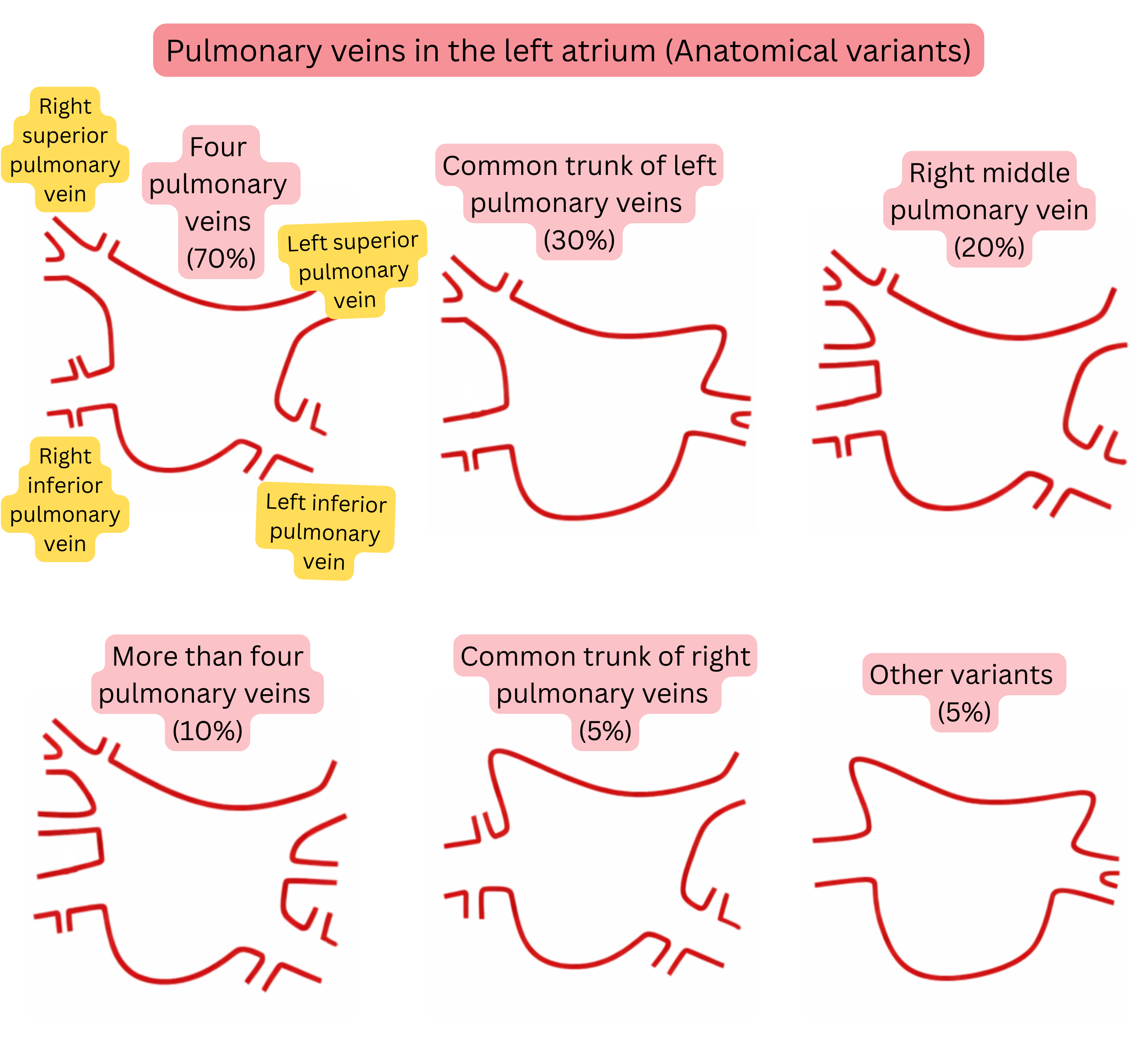
Anatomical Variants of Pulmonary Veins in the Left Atrium
Anatomical variant Prevalence
Typical anatomy (4 separate pulmonary veins) 60–70 %
Common trunk of the left pulmonary veins 20–30 %
Right middle pulmonary vein (accessory) 15–25 %
More than 4 pulmonary veins (accessory) 5–10 %
Common trunk of the right pulmonary veins <5 %
Early bifurcation of the pulmonary veins 10–15 %

Trigger and substrate

  • may be located in the same anatomical region, in close proximity, e.g. in the pulmonary vein ostia in the left atrium.
  • Alternatively, the trigger and substrate may be located in different regions, e.g. the trigger in the left atrial appendage and the substrate on the posterior wall of the left atrium.
Substrate of Atrial Fibrillation (Localization)
Anatomical site Basic anatomy Prevalence (%)
Posterior wall of the left atrium Bounded by the pulmonary veins. This is the most frequent site of fibrosis. 60–70 %
Pulmonary vein ostia Myocardial sleeves extending 1–4 cm into the pulmonary veins (especially the superior pulmonary veins) 50–60 %
Roof of the left atrium Region connecting the superior pulmonary veins. 30–40 %
Interatrial septum Region around the fossa ovalis and Bachmann’s bundle. 20–30 %
Left atrial appendage The trabeculated musculature of the appendage may represent a substrate. 10–20 %
Right atrium Substrate according to prevalence:
  • Crista terminalis (most common)
  • Lateral wall
  • Ostium of the superior vena cava
  • Ostium of the inferior vena cava
  • Ostium of the coronary sinus (least common)
10–20 %
Mitral isthmus Zone between the left inferior pulmonary vein and the mitral annulus. Critical for the development of perimitral flutter. 30–50% of atypical left atrial flutters pass through the mitral isthmus. 10–20 %

These guidelines are unofficial and do not represent formal guidelines issued by any professional cardiology society. They are intended for educational and informational purposes only.

Peter Blahut, MD

Peter Blahut, MD (Twitter(X), LinkedIn, PubMed)