Electrophysiology CINRE, hospital BORY
Atrial Fibrillation: Guidelines (2026) Compendium / 14.2 Pulsed Field Ablation of Atrial Fibrillation

Pulsed Field Ablation of Atrial Fibrillation


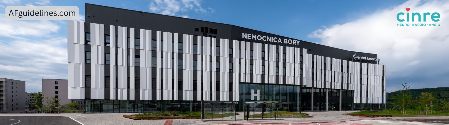
Before atrial fibrillation (AF) ablation, 4 weeks of anticoagulation therapy is recommended as prevention of thromboembolism.

  • Regardless of the CHA2DS2-VA score.
  • To minimize the risk of thrombus formation in the left atrial appendage as much as possible.
    • During ablation, manipulation of the catheter in the left atrium could dislodge a thrombus.
  • The risk of stroke in patients with AF and CHA2DS2-VA 0 is still approximately 0.5% (per year).

For prevention of thromboembolism, 2 months of anticoagulation therapy is recommended after ablation,

  • regardless of the CHA2DS2-VA score.
  • Electrical pulses cause endothelial injury of the left atrium,
    • subsequently activating the extrinsic coagulation pathway and a thrombus may form.
    • Endothelial healing after ablation takes approximately 2 months.
    • Therefore, anticoagulation therapy must be continued for 2 months after ablation regardless of the CHA2DS2-VA score.
  • After 2 months, anticoagulation therapy is continued according to the CHA2DS2-VA score.
Diagram of anticoagulation management in catheter ablation of atrial fibrillation illustrating the strategy before the procedure, immediately after ablation, during the first 2 months, and long-term decision-making based on the CHA₂DS₂-VA score.

Transoesophageal echocardiography before AF ablation (<24 h) is recommended despite 4 weeks of anticoagulation therapy in the following cases:

TEE before ablation – indications despite anticoagulation (4 weeks)
History of transient ischaemic attack (TIA)
History of stroke
Irregular use of anticoagulation therapy
INR < 2 (during warfarin therapy)
History of intracardiac thrombus (especially in the left atrial appendage)
History of left atrial appendage emptying velocity < 20 cm/s

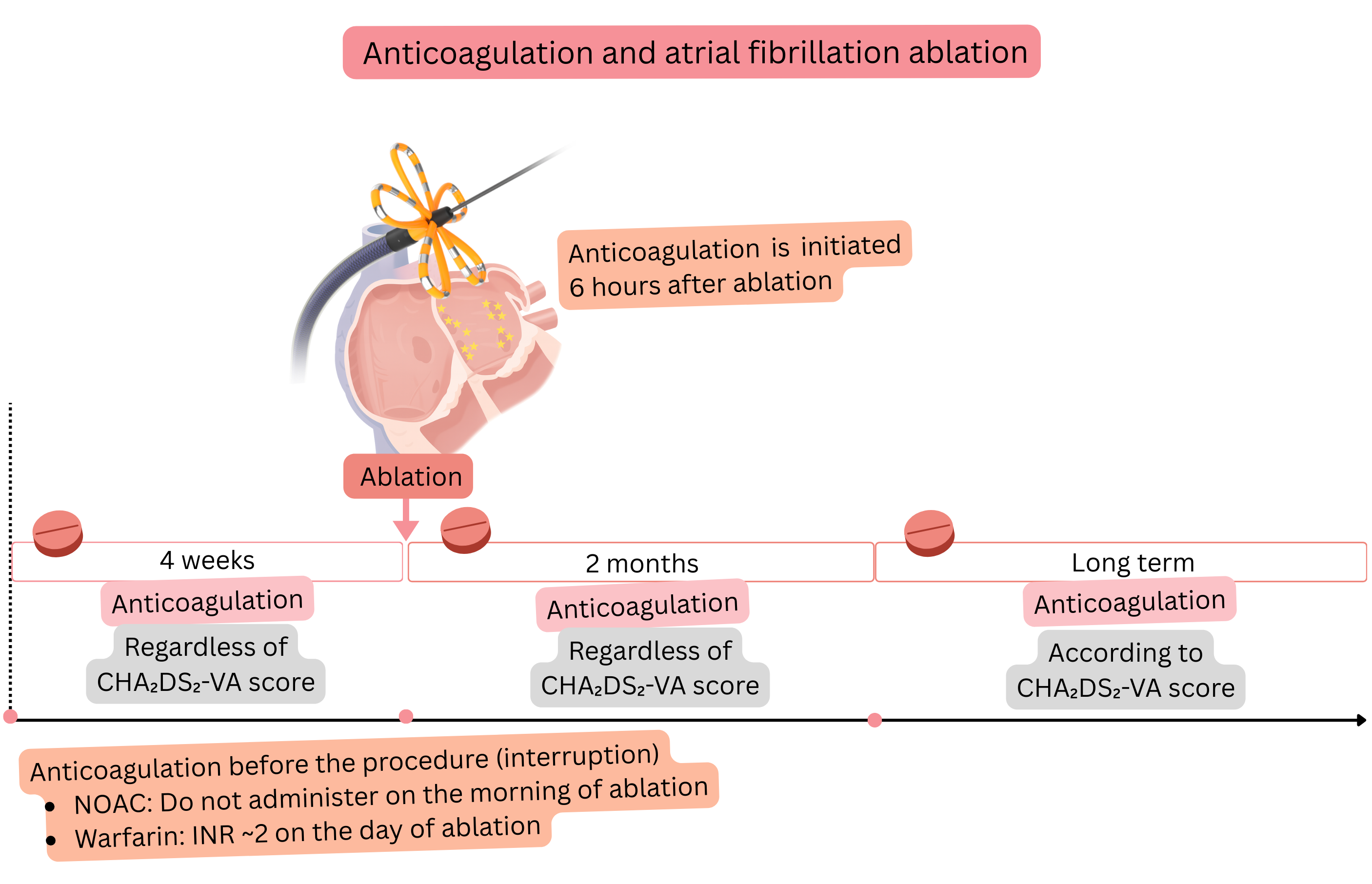
Before AF ablation, it is appropriate to discontinue anti-arrhythmic therapy (if the patient’s condition allows).

  • Because the therapy suppresses the arrhythmogenic substrate, which then becomes inactive during ablation and part of the substrate may remain untreated.
  • This suppressed substrate may not manifest at the end of ablation (during testing).

Before ablation, it is appropriate to discontinue anti-arrhythmic drugs used for rhythm control (if the patient’s condition allows), but not those used for rate control.

  • After AF ablation, procedural success is tested by atrial stimulation at 300/min. (simulation of a trigger),
  • if sustained AF (>1 min.) is induced during this stimulation, ablation is continued.
  • If the patient is taking anti-arrhythmic drugs for maintenance of sinus rhythm during ablation,
    • AF may not be inducible during testing and the ablation may be terminated.
    • However, the patient may develop AF, for example 3 months after ablation, once anti-arrhythmic therapy is completely discontinued.
Diagram of antiarrhythmic drug management in relation to catheter ablation of atrial fibrillation illustrating drug discontinuation before the procedure, preferred therapy during the first three months, and long-term strategy based on atrial fibrillation recurrence.
Discontinuation of anti-arrhythmic drugs before atrial fibrillation ablation
Drug Class Discontinuation before ablation
Disopyramide IA 3–5 days
Propafenone IC 3–5 days
Flecainide IC 3–5 days
Sotalol III 3–5 days
Amiodarone III 4–6 weeks
Dronedarone III 3–5 days

AF ablation (procedure) requires 3 vascular access sheaths via the groins (femoral veins):

  • Left groin – intracardiac echocardiography
  • Left groin – catheter advanced into the coronary sinus
  • Right groin – transseptal puncture needle and subsequently the Farawave catheter for pulsed field ablation

After insertion of the femoral sheaths, 5000 IU of unfractionated heparin (UFH) is administered.

  • The inserted sheaths and subsequently the catheters represent foreign material and activate the intrinsic coagulation pathway.
    • The extrinsic coagulation pathway is also partially activated due to vascular wall injury.
  • UFH is administered to prevent thrombus formation on the sheaths and catheters.
  • The intrinsic coagulation pathway is monitored using ACT or aPTT.
    • aPTT – a precise laboratory test, suitable for low doses of UFH. Normal range: 25–35 s
    • ACT – a rapid bedside test, suitable for high doses of UFH. Normal range: 80–120 s
  • The parameters (aPTT and ACT) assess the same coagulation pathway,
    • but are measured differently and therefore have different values.
      • aPTT is more precise at lower doses of UFH, e.g. in pulmonary embolism.
      • ACT is more precise and is used for immediate monitoring of high-level heparinization (UFH) during the procedure (AF ablation).
  • ACT prolongs within 1 min after UFH administration and begins to shorten after 20–30 min.
UFH dose and ACT prolongation
UFH dose ACT at 70 kg (prolongation from normal) ACT at 100 kg (prolongation from normal)
No UFH 80 – 120 s 80 – 120 s
1000 IU 120 – 140 s (↑20–40) 110 – 130 s (↑10–30)
3000 IU 150 – 180 s (↑50–80) 130 – 160 s (↑30–60)
5000 IU 200 – 240 s (↑100–140) 170 – 210 s (↑70–110)
7000 IU 230 – 280 s (↑130–180) 200 – 250 s (↑100–150)
10000 IU 280 – 340 s (↑180–240) 230 – 300 s (↑130–200)

A thrombus in the region of the venous sheaths and in the right atrium on the catheter is not as dangerous, as it embolizes to the lungs.

  • A thrombus in the left atrium is dangerous, as it may embolize to the brain and cause stroke.
Risk of thrombus formation on the sheath or catheter
ACT Estimated risk
80 – 120 s (no UFH) 10–20 % (within 10–20 min.)
250–300 s 1–2 %
300–350 s < 1 %

After introducing a dedicated needle via the femoral vein into the right atrium, a transseptal puncture is performed.

  • After the transseptal puncture, 3000 IU of UFH is administered again
    • and throughout the entire procedure an ACT of 300–350 s is maintained
  • A dedicated Farawave catheter is then advanced into the left atrium (where the pulmonary veins are located).
  • Pulmonary vein ablation (isolation) with pulsed field energy is then performed using the Farawave catheter.

ACT and UFH during the procedure

  • Throughout the entire procedure, an ACT of 300–350 s is maintained
  • ACT is checked every 20–30 min during the procedure (because after 20–30 min ACT begins to shorten)
  • According to the ACT value, 1000–3000 IU of UFH is added
  • ACT prolongs within 1 min after administration
Diagram of catheter ablation of atrial fibrillation illustrating isolation of the left superior pulmonary vein with the ablation catheter positioned in basket and flower configurations.

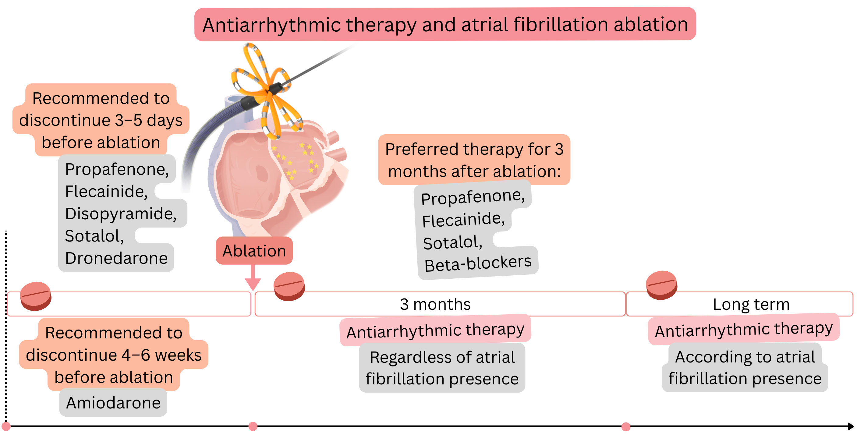
Farawave is a dedicated catheter with two configurations: basket and flower.

  • The Farawave catheter is advanced into the left superior pulmonary vein.
  • It is then set to the basket configuration and 2 electrical pulses are delivered; the catheter is then rotated by 20–30° and another 2 pulses are delivered.
  • The catheter is then set to the flower configuration and again 2 pulses are delivered; it is then rotated by 20–30° and another 2 pulses are delivered.
  • The pulmonary vein is then isolated; all 4 pulmonary veins are isolated sequentially in this manner.
  • Each vein requires at least 8 pulses for isolation.

3–5 min before the first application, 1 mg atropine is administered intravenously (may be repeated up to a maximum dose of 3 mg).

  • During delivery of electrical pulses in pulsed field AF ablation, the vagus nerve is stimulated,
    • with a risk of bradycardia or AV block.
  • If the patient is in AF during ablation and termination of AF with conversion to sinus rhythm occurs during pulsing,
    • post-conversion bradycardia may occur.
Atropine – prevention of bradycardia before ablation
Purpose Prevention of bradycardia
Mechanism Blocks vagal effect (antimuscarinic effect)
Dose 1 mg intravenous (max. 3 mg)
Onset of action 1–2 min
Expected effect Heart rate increase by 20–40/min
Duration of action 30–60 min
Contraindication Glaucoma

Electrical pulses are painful; therefore, they are delivered under general anaesthesia or sedation.

If AF persists after pulmonary vein isolation, a more extensive left atrial ablation is performed:

  • Posterior wall → mitral isthmus, until AF terminates (if the patient was in AF before ablation).
  • If AF does not terminate despite extensive ablation, electrical cardioversion is performed.

Left atrial appendage and ablation.

  • Ablation of the left atrial roof is not recommended, because it disrupts Bachmann’s bundle,
    • which also innervates the left atrial appendage.
    • The appendage then loses its physiological mechanical function and thrombus may form within it.
  • Ablation at the left atrial appendage ostium is not recommended,
    • because it electrically isolates the appendage and it loses mechanical function.
    • The appendage then loses its physiological mechanical function and thrombus may form within it.

If a patient has AF and atrial flutter (AFL),

  • in the same session, ablation of both AF and the cavotricuspid isthmus using the Farawave catheter is recommended.

During pulsing in AF and AFL ablation, coronary spasm may occur if ablation is performed near a coronary artery.

  • In the region of the mitral isthmus lies the circumflex artery,
  • in the region of the tricuspid isthmus lies the right coronary artery.
  • Before isthmus ablation, nitroglycerin or Isoket is administered to prevent coronary spasm.
  • If ST elevation occurs during isthmus ablation, an additional dose is recommended.
Prevention of coronary spasm before ablation – Isoket vs. nitroglycerin
Drug Isoket (isosorbide dinitrate) Nitroglycerin
Purpose Slower, longer-lasting prevention of coronary spasm Rapid prevention and treatment of coronary spasm
Mechanism of action NO donor → coronary vasodilation NO donor → coronary vasodilation
Administration intravenous bolus 1–2 mg intravenous bolus 50–200 µg
Onset of action 3–5 min 1–2 min
Duration of action 30–60 min 5–10 min
Risk of hypotension Higher (longer duration) Lower (short duration)

ECG changes during ablation in the region of the mitral and tricuspid isthmus:

  • Mitral isthmus – ST elevation may occur (I, aVL, V5–6)
  • Cavotricuspid isthmus – ST elevation may occur (II, III, aVF)

Electrical pulses in pulsed field ablation are cardioselective:

  • They cause selective electroporation followed by apoptosis of cardiomyocytes only (where the arrhythmogenic foci and AF substrate are located),
  • The electrical pulses do not injure surrounding tissues (veins, oesophagus, nerves).
Electrical pulses in pulsed field ablation
Do not cause pulmonary vein stenosis
Do not cause atrio-oesophageal fistula
Do not cause phrenic nerve injury

After AF ablation, procedural success can be verified in three ways:

  1. Pacing from the pulmonary veins
    • The Farawave catheter is advanced into the pulmonary vein in the “basket” configuration,
    • then from each spline a train of stimuli (6–8 stimuli) is delivered with a cycle length of 500 ms.
    • Atrial response is assessed on the catheter positioned in the coronary sinus (CS),
      • if signals in the vein are independent of signals on the CS catheter → the vein is isolated,
      • if signals on the CS catheter regularly follow pacing from the vein → the vein is not isolated.
    • Pacing from the pulmonary veins tests only pulmonary vein isolation,
      • it does not test arrhythmogenic substrate outside the pulmonary vein ostia.
  2. Burst pacing
    • Pacing is performed from the catheter positioned in the coronary sinus,
      • the principle is that stimuli propagate to the left atrium.
    • A burst is a train of impulses consisting of 8–10 stimuli with the same cycle length (e.g. 300 ms).
    • It starts with a burst at 300 ms,
      • and the cycle length of each subsequent train is progressively reduced by 10–30 ms,
      • with a pause of 5–10 s between trains,
      • the train cycle length is reduced down to atrial refractoriness
        • or to a cycle length of 200 ms, corresponding to a rate of 300/min.
    • If a substrate for AF is present in the left atrium, burst pacing induces AF.
    • Ablation is unsuccessful if burst pacing induces AF lasting >1 min.
      • if AF terminates spontaneously within 1 min, ablation is considered successful.
  3. Ramp pacing
    • Pacing is performed from the catheter positioned in the coronary sinus,
      • the principle is that stimuli propagate to the left atrium.
    • Continuous pacing without pauses is performed, with the cycle length progressively shortened by 10–20 ms.
      • It starts at 300 ms and the cycle length is continuously shortened after 3–5 stimuli.
    • The ramp is continued to a cycle length of 200 ms or to atrial refractoriness.
    • If a substrate for AF is present in the left atrium, ramp pacing induces AF.
    • Ablation is unsuccessful if ramp pacing induces AF lasting >1 min.
      • if AF terminates spontaneously within 1 min, ablation is considered successful.

Verification of atrial fibrillation ablation success
Method Site of pacing What is assessed Success
Pacing from the pulmonary veins Sequentially from each pulmonary vein (using the Farawave catheter) Whether impulses from the vein propagate to the left atrium No atrial response = vein isolated
Burst pacing From the coronary sinus (CS catheter) Induction of AF after bursts (8–10 stimuli) AF is not induced or AF does not last > 1 min = success
Ramp pacing From the coronary sinus (CS catheter) Whether AF is induced during pacing with progressive cycle shortening AF is not induced or AF does not last > 1 min = success

During pulsed field ablation, electroporation of cardiomyocytes occurs, which is perforation of the cell membrane by an electric field. This is followed by apoptosis. Cardio-specific enzymes are released from damaged cardiomyocytes and rise to high levels within 24 h.

Cardio-specific enzymes within 24 h after pulsed field ablation
Enzyme Value
Troponin 1500 ng/l (±500)
CK 300 ng/l (±100)
CK-MB 35 mg/l (±10)

During pulsed field ablation, haemolysis occurs, which manifests after the procedure as macroscopic haematuria.

  • Acute kidney injury may occur; therefore, serum creatinine is checked after the procedure.
  • Acute kidney injury (definition):
    • Increase in creatinine by ≥26.5 µmol/L (≥0.3 mg/dL) within 48 h or
    • Increase in creatinine to ≥1.5-fold of baseline within 7 days
  • As prevention of acute kidney injury, the patient is hydrated after ablation; it is recommended to administer:
    • 2000 ml of normal saline over 24 h.
  • The risk of acute kidney injury is <1%.

Ablation success is assessed by the presence of AF recurrence within 1 year after ablation.

  • Success is higher in paroxysmal AF, because paroxysmal AF is almost always localized to the pulmonary vein ostia.
  • Persistent AF is also localized outside the pulmonary veins and these substrates may not always be successfully ablated.
Atrial fibrillation ablation success (within 12 months)
AF classification Success
Paroxysmal AF 66–82 %
Persistent AF 56–72 %

Blanking period

  • After pulsed field ablation, atrial myocardial electroporation, endothelial injury, local inflammation, and oedema occur.
    • In this setting, conduction disturbances and atrial arrhythmias, including AF, may occur.
  • The blanking period is the time interval—the first 3 months after AF ablation,
    • during which episodes of atrial arrhythmia (including AF) are not considered AF recurrence.
  • Therefore, during the blanking period (the first 3 months after ablation), the following are always prescribed:
    • Anti-arrhythmic drugs (first 3 months), because arrhythmogenic risk in the ablation lesion persists for approximately 3 months
    • Anticoagulation therapy (first 2 months), because thrombotic risk in the ablation lesion persists for approximately 2 months.
Diagram of the blanking period after catheter ablation of atrial fibrillation illustrating antiarrhythmic and anticoagulation strategies during the first three months and subsequent long-term management based on atrial fibrillation recurrence and CHA₂DS₂-VASc score.
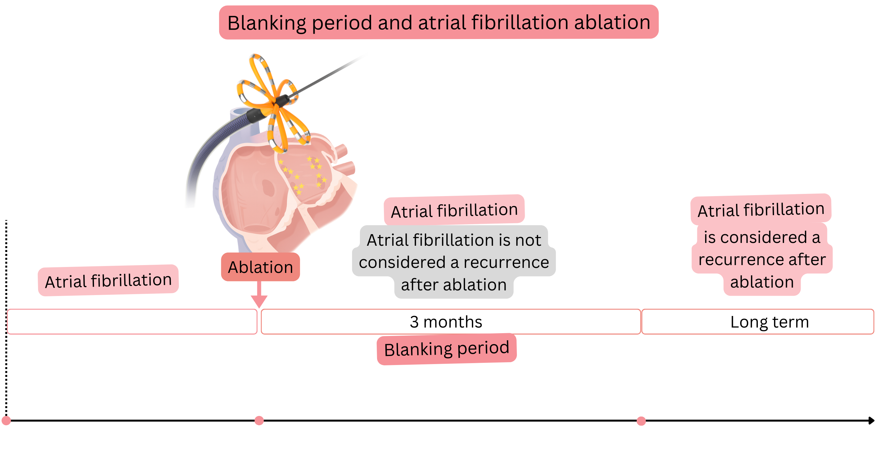
Therapy during the blanking period (3 months after ablation)
Therapy Duration Note
Anti-arrhythmic drugs 3 months Administered regardless of whether sinus rhythm or AF is present.
Anticoagulation therapy 2 months Administered regardless of the CHA2DS2-VA score

In AF recurrence after ablation—pulmonary vein isolation (with pulsed field energy), reconnection of a pulmonary vein to the left atrium may occur. Reconnection most commonly occurs in:

  • Right inferior pulmonary vein – 42 %
  • Right superior pulmonary vein – 35 %
  • Left superior pulmonary vein – 27 %
  • Left inferior pulmonary vein – 19 %

During and after pulsed field AF ablation, complications may occur, but they are very rare. The incidence of complications is:

  • Major complications (0.98 %)
  • Minor complications (3.21 %)

Major and minor complications are listed in the following table:

Major complications of pulsed field ablation
Total 0.98 %
Pericardial tamponade 0.36 %
Vascular complication (requiring intervention) 0.30 %
Coronary spasm 0.14 %
Stroke 0.12 %
Haemolysis with acute renal failure 0.03 %
Death 0.03 %
Other (thrombosis, coronary air embolism) 0.006 %
Oesophageal fistula 0 %
Pulmonary vein stenosis 0 %
Phrenic nerve injury (permanent) 0 %

Minor complications of pulsed field ablation
Total 3.21 %
Vascular complications (not requiring intervention) 2.20 %
Pericardial effusion (not requiring intervention) 0.33 %
Other minor complications (haematomas, arrhythmias) 0.32 %
Pericarditis 0.17 %
Transient ischaemic attack 0.12 %
Phrenic nerve injury (temporary) 0.06 %

In 5–15% of patients, asymptomatic silent cerebral ischaemia occurs during pulsed field ablation.

  • It occurs due to microembolization (microthrombi, air bubbles, microparticles from the left atrial endothelium).
  • Microemboli are released during delivery of electrical pulses.
  • In the future, impairment or worsening of cognitive function (attention, memory, learning) may occur.
Atrial fibrillation ablation Class
Pulsed field ablation (not radiofrequency or cryoablation) is recommended as the preferred method of atrial fibrillation ablation. I
Pulsed field ablation is recommended in patients with paroxysmal or persistent atrial fibrillation if atrial fibrillation is symptomatic:
  • despite optimized anti-arrhythmic therapy or
  • if anti-arrhythmic therapy is not possible due to adverse effects or contraindications
I
Pulsed field ablation is recommended in patients with tachycardia-induced cardiomyopathy due to atrial fibrillation. I
Pulsed field ablation should be considered in patients with atrial fibrillation who have symptomatic pre-automatic pauses. IIa
In atrial fibrillation recurrence, pulsed field ablation may be repeated (not earlier than 3 months) if atrial fibrillation is symptomatic:
  • despite optimized anti-arrhythmic therapy or
  • if anti-arrhythmic therapy is not possible due to adverse effects or contraindications
IIa
Before atrial fibrillation ablation, CT or MR angiography of the left atrium and pulmonary veins should be considered to assess pulmonary vein anatomy. IIa
The “pace and ablate” strategy may be considered in patients with symptomatic atrial fibrillation in whom the following have failed:
  • pharmacological therapy and
  • ≥2 ablations (pulsed field)
IIa
Anticoagulation therapy and atrial fibrillation ablation Class
Anticoagulation therapy is recommended for at least 4 weeks before atrial fibrillation ablation, regardless of the CHA₂DS₂-VA score. I
NOAC anticoagulation therapy is recommended not to be taken in the morning on the day of atrial fibrillation ablation. I
NOAC anticoagulation therapy is recommended to be started 6 h after atrial fibrillation ablation if there are no signs of bleeding. I
During warfarin therapy, atrial fibrillation ablation is recommended to be performed with a therapeutic INR of approximately 2.0 on the day of the procedure. I
Anticoagulation therapy is recommended for the first 2 months after atrial fibrillation ablation, regardless of ablation success and regardless of the CHA₂DS₂-VA score. I
Two months after atrial fibrillation ablation, long-term anticoagulation is indicated according to the CHA₂DS₂-VA score, regardless of ablation success. I
Anti-arrhythmic therapy (propafenone, flecainide, sotalol, beta-blockers) is recommended for the first 3 months after atrial fibrillation ablation, regardless of ablation success. I
Three months after atrial fibrillation ablation, anti-arrhythmic therapy is indicated according to atrial fibrillation recurrence. I
Atrial fibrillation ablation may be considered if the patient is receiving dual antithrombotic therapy (e.g. NOAC + clopidogrel). IIa
Atrial fibrillation ablation during cardiac surgery Class
In a patient undergoing cardiac surgery on the mitral valve, concomitant surgical atrial fibrillation ablation using the Cox-Maze IV procedure is recommended. I
In a patient undergoing cardiac surgery other than mitral valve surgery, concomitant surgical atrial fibrillation ablation using the Cox-Maze IV procedure should be considered. IIa
During cardiac surgery, the presence of thrombus in the left atrium is recommended to be excluded before surgical atrial fibrillation ablation. I

These guidelines are unofficial and do not represent formal guidelines issued by any professional cardiology society. They are intended for educational and informational purposes only.

Peter Blahut, MD

Peter Blahut, MD (Twitter(X), LinkedIn, PubMed)