Electrophysiology CINRE, hospital BORY
Atrial Fibrillation: Guidelines (2026) Compendium / 7.4.4 Hypertrophic Cardiomyopathy and Atrial Fibrillation

Hypertrophic Cardiomyopathy and Atrial Fibrillation


Hypertrophic cardiomyopathy (HCM) is a congenital genetic disease,

  • characterized by left ventricular hypertrophy and subsequent diastolic dysfunction.
  • Progressive atrial remodeling and dilatation occur, creating a substrate for atrial fibrillation (AF).
Illustration of atrial fibrillation in hypertrophic cardiomyopathy with echocardiographic evidence of left ventricular hypertrophy with wall thickness >15 mm and indication for anticoagulation with NOACs or warfarin.

AF and HCM are associated with a high thromboembolic risk,

  • therefore, this condition requires anticoagulant therapy regardless of the CHA2DS2-VA score.

The prevalence of HCM in the population is 1/500.

AF occurs in 20–40% of patients with HCM.

HCM with AF carries a thromboembolic risk of 5–10% per year

  • in patients without anticoagulant therapy

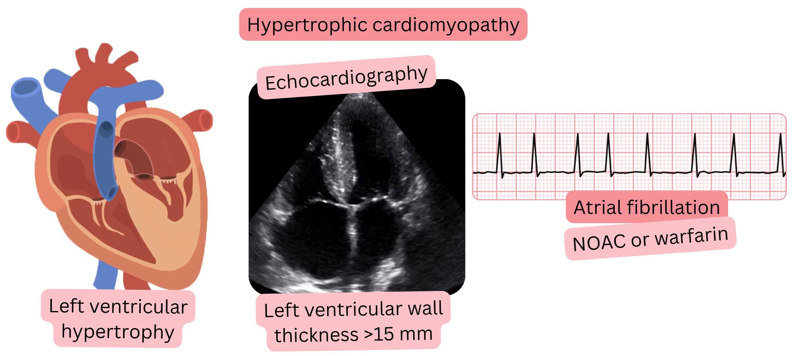
Echocardiography and HCM:

  • Left ventricular wall thickness ≥ 15 mm in any segment of the left ventricle.
Anticoagulant Therapy and Hypertrophic Cardiomyopathy Class
In patients with hypertrophic cardiomyopathy and atrial fibrillation, anticoagulant therapy (preferably NOAC) is indicated regardless of the CHA2DS2-VA score. I

Hypertrophic cardiomyopathy produces characteristic findings on ECG and transthoracic echocardiography, summarized in the table below.

The following are used for definitive diagnosis of hypertrophic cardiomyopathy:

  • Cardiac MRI and genetic testing
Hypertrophic Cardiomyopathy – ECG and Transthoracic Echocardiography (TTE)
Investigation Characteristic findings
ECG
  • High QRS voltage (LVH criteria):
    • Sokolow–Lyon index: S(V1) + R(V5/V6) ≥ 35 mm
    • Cornell index: R(aVL) + S(V3) ≥ 28 mm (men), ≥ 20 mm (women)
  • Deep negative T waves
  • Septal or inferior Q waves
  • Repolarization abnormalities
  • Supraventricular and ventricular arrhythmias
TTE
  • Asymmetric septal hypertrophy
  • Left ventricular hypertrophy ≥ 15 mm (any segment of the left ventricle)
  • SAM (Systolic Anterior Motion) phenomenon
    • systolic displacement of the anterior mitral leaflet
    • towards the hypertrophied septum, resulting in:
      • mitral regurgitation and LVOT obstruction
  • LVOT obstruction (gradient > 30 mmHg)

LVOT - Left Ventricular Outflow Tract


These guidelines are unofficial and do not represent formal guidelines issued by any professional cardiology society. They are intended for educational and informational purposes only.

Peter Blahut, MD

Peter Blahut, MD (Twitter(X), LinkedIn, PubMed)