Electrophysiology CINRE, hospital BORY
Atrial Fibrillation: Guidelines (2026) Compendium / 7.4.3 Cardiac Amyloidosis and Atrial Fibrillation

Cardiac Amyloidosis and Atrial Fibrillation


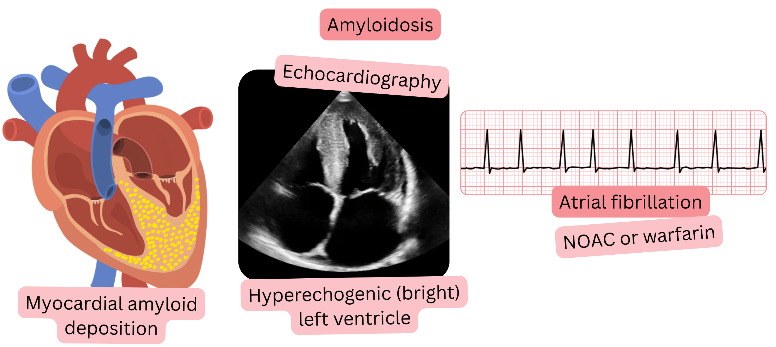
Amyloidosis is a systemic infiltrative disease in which amyloid is deposited in the extracellular space of the myocardium; this condition is referred to as cardiac amyloidosis.

Illustration of atrial fibrillation in cardiac amyloidosis with a hyperechogenic left ventricle on echocardiography and indication for anticoagulation with NOACs or warfarin.

Cardiac amyloidosis

  • Leads to ventricular hypertrophy without dilatation and to diastolic dysfunction due to “stiff ventricles”,
    • resulting in restrictive cardiomyopathy and subsequent atrial dilatation.
  • Infiltration of the valves frequently results in aortic stenosis.
  • Cardiac amyloidosis with atrial fibrillation (AF) represents a high risk of thromboembolism,
    • because the atria are dilated and remodeled due to amyloid deposits.

Amyloidosis may be

  • Hereditary, which is very rare (< 1%)
  • Acquired (senile)
    • Present in 10–20% of patients > 65 years with HFpEF (heart failure with preserved ejection fraction)
    • At older age, due to altered protein synthesis, the liver begins to produce amyloid

Aortic stenosis is present in 10–20% of patients with cardiac amyloidosis.

AF is present in 50–70% of patients with cardiac amyloidosis.

Cardiac amyloidosis with AF carries a thromboembolic risk of 5–10% per year

  • in patients without anticoagulant therapy
Anticoagulant Therapy and Cardiac Amyloidosis Class
In patients with cardiac amyloidosis and atrial fibrillation, anticoagulant therapy (preferably NOAC) is always indicated, regardless of the CHA2DS2-VA score. I

Cardiac amyloidosis produces characteristic findings on ECG and transthoracic echocardiography, which are summarized in the table below.

The following modalities are used for definitive diagnosis of cardiac amyloidosis:

  • Cardiac MRI, cardiac scintigraphy, and endomyocardial biopsy
Cardiac Amyloidosis – ECG and Transthoracic Echocardiography (TTE)
Investigation Characteristic findings
ECG
  • Low QRS voltage (< 5 mm in limb leads, < 10 mm in precordial leads)
  • Pseudoinfarction pattern with Q waves (in precordial or inferior leads)
    • without a history of myocardial infarction
  • Conduction system abnormalities:
    • AV block I–III degree
    • Bundle branch block
    • Intraventricular conduction delay
TTE
  • Concentric left ventricular hypertrophy
  • HFpEF (Heart Failure with Preserved Ejection Fraction)
  • Atrial dilatation
  • Hyperechogenic hypertrophied ventricles (amyloid increases echogenicity)

These guidelines are unofficial and do not represent formal guidelines issued by any professional cardiology society. They are intended for educational and informational purposes only.

Peter Blahut, MD

Peter Blahut, MD (Twitter(X), LinkedIn, PubMed)