Electrophysiology CINRE, hospital BORY
Atrial Fibrillation: Guidelines (2026) Compendium / 2.2 Pathophysiology of Atrial Fibrillation

Pathophysiology of Atrial Fibrillation


The substrate (trigger and substrate) for atrial fibrillation (AF) develops when an electrical disorder of the atria occurs as a result of atrial remodelling (structural, mechanical, or electrical). Atrial remodelling may be:

  • Acute (within several hours)
  • Chronic (over several years)

Acute atrial remodelling

  • Develops within several hours during an acute critical condition (e.g. myocardial infarction).
  • Acute remodelling often resolves after treatment of the critical condition.
  • Acute remodelling is usually reversible.

Chronic atrial remodelling

  • Develops over several years due to atrial cardiomyopathy (ACMP).
    • 10% develop spontaneously without risk factors (genetic ACMP).
    • 90% develop due to risk factors.
  • Chronic remodelling is irreversible.

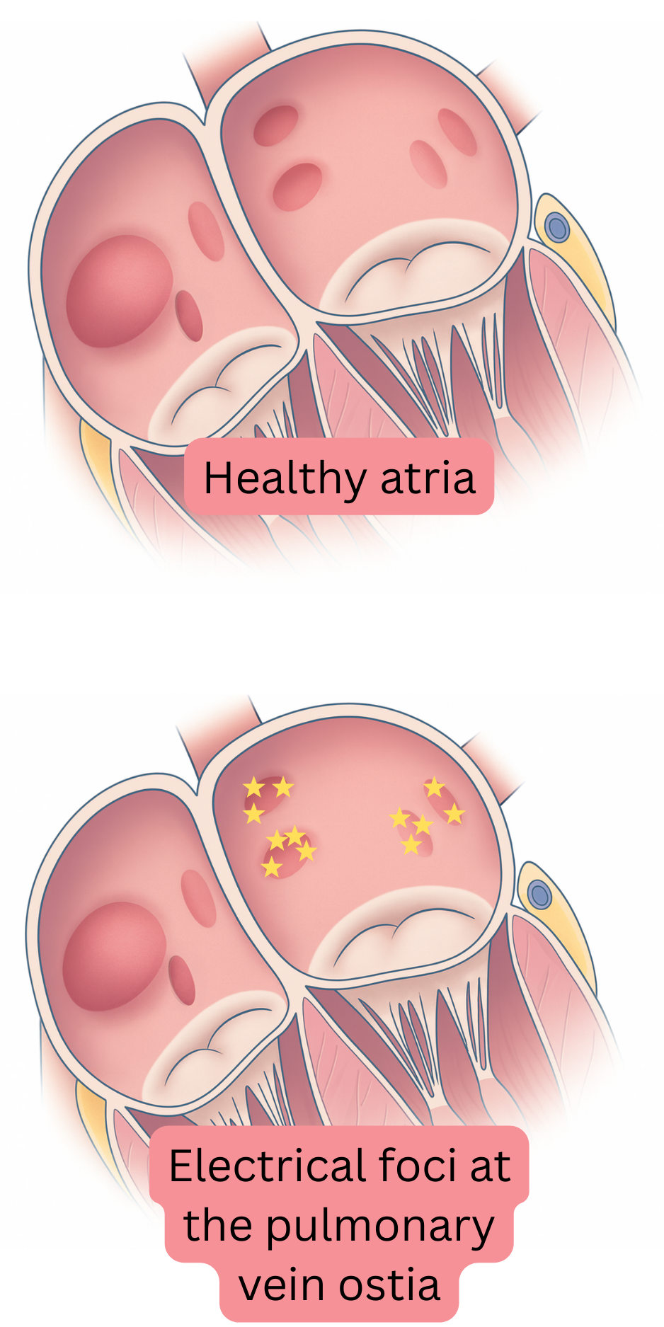
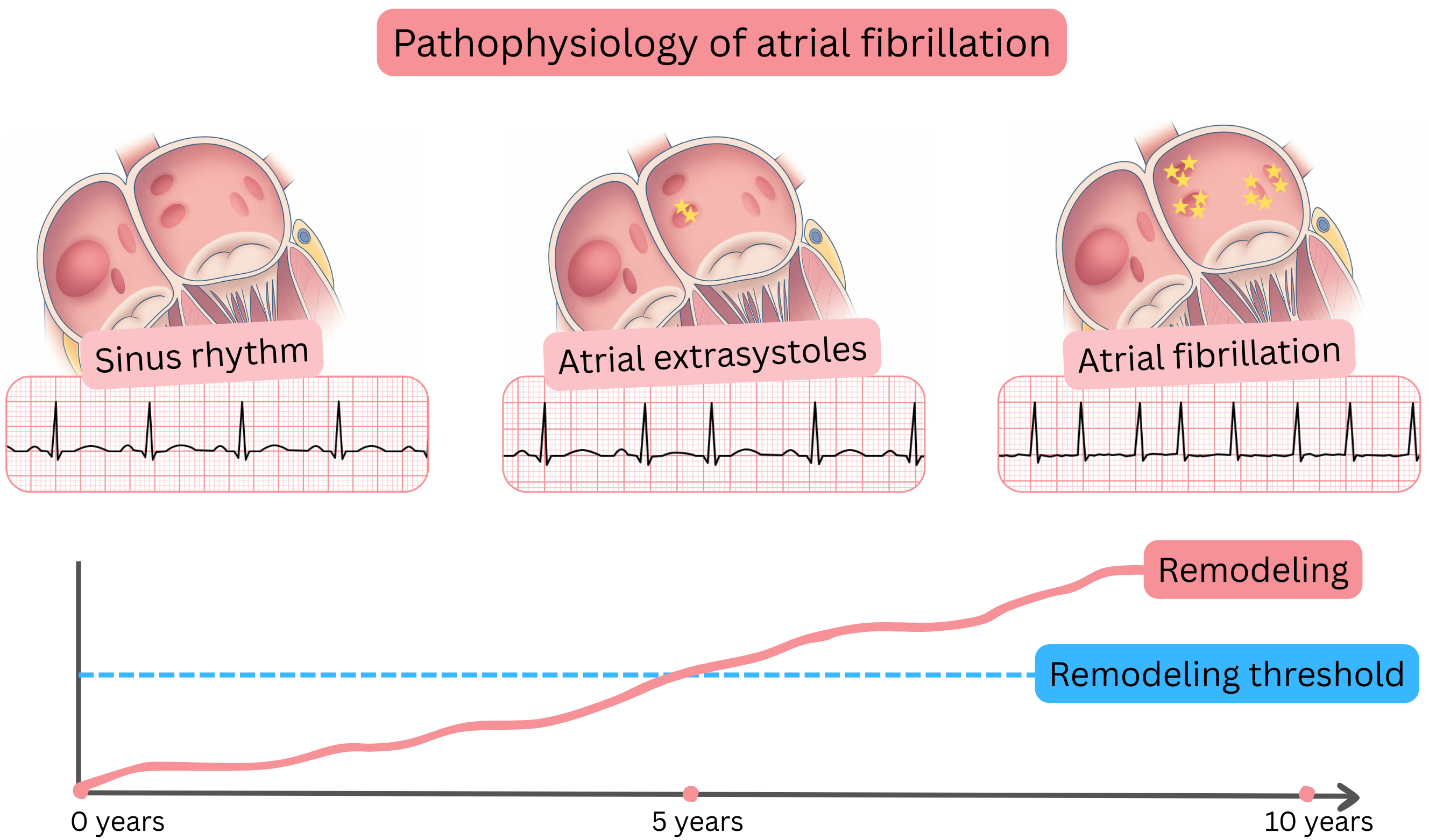
Atrial remodelling leads to the development of a trigger in the atria, with multiple electrical foci (5–20) generating impulses independently and activating the substrate. The resulting impulse rate is 300–600/min. The atria no longer produce synchronous systole but instead fibrillate—“quiver”.

Remodelling most commonly begins in the region of the pulmonary vein ostia in the left atrium, where both the trigger and substrate for AF gradually develop. The ostium is the anatomical region where the pulmonary vein connects to the left atrium. Remodelling then progressively extends to the entire left atrium and partially to the right atrium over 5–15 years.

Illustration comparing healthy atria with electrical foci at the pulmonary vein ostia acting as triggers of atrial fibrillation.
Pathophysiology of atrial fibrillation
The substrate (trigger and substrate) for atrial fibrillation most commonly develops due to atrial cardiomyopathy.
The pre-fibrillatory state consists of premature atrial contractions or atrial tachycardia originating from the region of the pulmonary vein ostia, which may sometimes be visible on the ECG.
The pre-fibrillatory state progresses within 6–12 months to paroxysmal atrial fibrillation.
Paroxysmal atrial fibrillation is most commonly triggered by premature atrial contractions or atrial tachycardia originating from the pulmonary vein ostia.

The pulmonary vein ostia have different electrophysiological properties compared with the atrial myocardium. The ostia contain the highest density of autonomic nerve fibres and have a short refractory period. Therefore, even minimal remodelling in the ostial region readily leads to the development of foci (triggers) that generate impulses through triggered activity or abnormal automaticity. These foci then generate premature atrial contractions or atrial tachycardia, which activate the substrate and initiate AF.

Pre-fibrillatory state

  • In remodelled pulmonary vein ostia, premature atrial contractions (triggers) or short-lasting (<60 s) atrial tachycardia initially develop. These arrhythmias can very rarely be captured on ECG or Holter monitoring.
  • This represents the so-called pre-fibrillatory state; the patient does not yet have AF, but in the near future (within 6–12 months) the left atrium reaches the so-called remodelling threshold and AF develops.

Remodelling threshold

  • If remodelling exceeds the remodelling threshold (substrate develops), AF occurs.
  • AF develops when premature atrial contractions or atrial tachycardia from the pulmonary vein ostia (triggers) activate foci in the ostia and in the left atrial myocardium (substrate). These foci then generate impulses at a rate of 300–600/min.
  • AF most commonly begins as paroxysmal AF (spontaneously terminates within 7 days).
Diagram of atrial fibrillation pathophysiology illustrating progression from sinus rhythm through atrial ectopy to atrial fibrillation with progressive atrial remodeling over time.

Trigger of atrial fibrillation

  • AF is most commonly triggered by premature atrial contractions or atrial tachycardia.
  • Premature beats or tachycardia most commonly originate from foci in the pulmonary vein ostia, although foci may also be located outside the ostial region.

Episode of atrial fibrillation

  • During an episode of AF, impulses arise in electrical foci (within the substrate) independently at a rate of 300–600/min. The SA node is deactivated because it is continuously depolarized by impulses from these foci.
  • The atria depolarize asynchronously and fibrillate in an uncoordinated manner at a rate of 300–600/min.
  • Atrial electrical activity is transmitted irregularly to the ventricles through the AV node, which acts as a filter, most commonly with a ventricular rate of <100/min.
Diagram of preexcited atrial fibrillation with a short SPERRI below 250 ms indicating a malignant accessory pathway and rapid ventricular responses on ECG.

Pre-excited atrial fibrillation

  • If the patient has an antegrade accessory pathway, impulses during AF are conducted to the ventricles both via the accessory pathway and via the AV node.
  • Pre-excited AF means that during AF the ECG shows a delta wave.
  • Such a patient must not receive drugs that block the AV node, as ventricular fibrillation may occur.
  • Ventricular fibrillation may occur if the patient has a malignant accessory pathway (SPERRI <250 ms).

These guidelines are unofficial and do not represent formal guidelines issued by any professional cardiology society. They are intended for educational and informational purposes only.

Peter Blahut, MD

Peter Blahut, MD (Twitter(X), LinkedIn, PubMed)